Connectivity-based parcellation atlases |
In a few recent publications, we investigated whether it is possible to subdivide parts of the human cerebral cortex based on their connections with the rest of the brain. Here we present the results of these parcellations in the form of atlases for FSLview, part of the popular MRI analysis software package FSL. To install any of the atlases, download the compressed volume below and unpack it into the directory /data/atlases of your FSL version. The atlas should then be accessible in FSLview in the same way as other atlases are. Note that you will be using these atlases strictly at your own risk. The current versions of all atlases are made available purely as a pilot project. Currently, we have atlases available of lateral parietal cortex (Mars et al., 2011, J Neurosci), the temporoparietal junction area (TPJ) (Mars et al., 2012, Cereb Cortex), the dorsal frontal cortex (Sallet et al., 2013, J Neurosci), the ventral frontal cortex (Neubert et al., 2014, Neuron), and the cingulate and orbitofrontal cortex (Neubert et al., 2015, PNAS). All atlases were created as follows. The images of each mask for each participant in the studies were converted to MNI152 2mm space, dilated when necessary, binarized, and added together. The resulting images were thresholded at 25% of participants, so only voxels that belong to any given mask in 25-100% of the participants in a study will be labelled. The summary image shows images thresholded at 50% or 75% of participants. When you press the "Locate selected structure" button FSLview will guide you to the voxel that was chosen as representative of that cluster in its respective paper. This is usually the centre of gravity of the mask, which explains why sometimes the voxel might fall outside the direct grey matter boundary. The MNI152 1mm atlas files were created by resampling all files in that resolution. When you use these atlases, please consider referencing the appropriate papers describing the parcellation study. We'd very much like to receive your feedback. Please send an e-mail. Parietal cortex
In our parietal parcellation study we subdivided both the superior and inferior parietal lobule into subregions, which we subsequently compared with previous parcellations based on cytoarchitecture and with known subdivisions in the macaque brain. We identified five areas in the superior parietal cortex and five regions in the inferior parietal lobule. We refer to these regions as follows. The inferior parietal lobule regions are labeled anterior to posteriorly as IPLA (red in Fig. 2 of the paper), IPLB (blue), IPLC (green), IPLD (pink), and IPLE (yellow). The superior parietal lobule regions are labelled SPLA (red in Fig. 7 of the paper), SPLB (blue), SPLC (green), SPLD (pink), and SPLE (yellow). Note that the parcellation was performed on the right hemisphere only.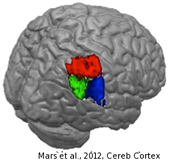 Temporoparietal junction area (TPJ)
In our temporoparietal junction area (TPJ) parcellation study we tested the hypothesis that the large cortical expanse referred to in the literature as "TPJ" consist of different subdivisions that participate in different cortical networks. We found that TPJ can be subdivided into (at least) three subdivisions. A dorsal subdivision (right, red) overlaps with the inferior parietal lobule that was further subdivided in the parietal study described above. Two ventral subdivisions, which we refer to as TPJa and TPJp (right, blue and green), show similarities with TPJ as identified in the attentional and social literature, respectively. Note that the parcellation was performed on the right hemisphere only.Dorsal frontal cortex
|

 In Franz-Xaver Neubert's
|